
CASE STUDY · KERATOCONUS
A refraction that would not repeat
A man in his seventies, forty years in small-diameter rigid lenses, referred for spectacles. The lenses correcting his cornea had been reshaping it.
Why he was referred
He came to us for a second opinion on glasses, not on lenses. He had worn small-diameter corneal rigid gas permeable lenses for more than forty years for keratoconus, was entirely dependent on them, and could not function in spectacles because of glare and distortion. Strabismus surgery was scheduled, and part of the reason for the referral was to give him usable glasses for the post-operative period.
The problem showed up in the refraction itself. His subjective responses would not repeat from one presentation to the next — not merely difficult, but inconsistent in a way that kept changing the endpoint.
That pattern is the finding. Keratoconus produces irregular astigmatism; it does not ordinarily produce refractive instability of that character. An unreliable subjective refraction in a long-term rigid lens wearer should raise corneal warpage before the refraction is written off as a hard one.
What examination showed
Anterior segment imaging and slit lamp examination demonstrated significant bilateral corneal warpage consistent with chronic rigid lens bearing. There was also a large subepithelial nodule on the left cornea with the appearance of Salzmann’s nodular degeneration, sitting immediately adjacent to the nasal edge of the lens landing zone.
Two things followed from that. The device correcting his cornea was actively moulding it, so any measurement taken that day described a shape that would not persist. And the nodule lay exactly where the lens bore down, which is the pattern of a mechanically provoked lesion rather than a primary one.
The decision: fitting was deliberately deferred
A scleral lens ordered on that first measurement would have been built to a transient cornea. So rigid lens wear was discontinued for five weeks as a planned washout before any diagnostic lens was applied. The strabismus surgery made the interval convenient, but the lens fitting was withheld on corneal grounds, not scheduling ones.
At five weeks, measurable regression had occurred: about a line of acuity in each eye, a documented shift in both the magnitude and the axis of the astigmatism bilaterally, and near-complete regression of the Salzmann’s nodule without any surgical intervention.
On the interval itself, honestly: published series report a mean time to refractive stability of roughly nine weeks in rigid lens wearers, with a range extending much longer, and a common clinical rule of thumb allows a month out of lenses for each decade of wear. Against either benchmark, five weeks was short for forty years of wear. It was markedly better than none and probably shorter than ideal — and the two early lens remakes described below are at least as consistent with a cornea still settling as with anything else.
The fitting
Diagnostic scleral lenses of 17.0 mm were applied, a diameter chosen to clear both cornea and limbus given horizontal visible iris diameters of 12.40 mm and 12.29 mm. Anterior segment OCT over the settled lenses confirmed complete corneal vault and 360° limbal clearance in both eyes, with measured clearances of 255–346 µm on the right and 210–285 µm on the left.
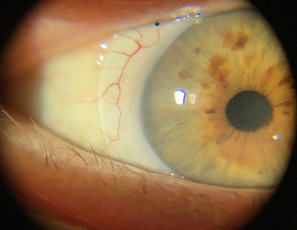
His lens in place — the edge crossing the sclera below the limbus, clear of the cornea entirely.
The lens finally retained was a back-surface and front-surface toric design with an asymmetric periphery, 7.00 mm base curve, in a high-Dk material. The figure that mattered was the 450 µm difference between the horizontal and vertical sagittal depths. The first order carried a 300 µm differential; the lens that worked needed half as much again, which matches the rotational instability and decentration documented during the fitting.
What the follow-up course actually involved
At dispense he read 20/20 in each eye and said he had not seen that well in forty years. What came after is the part usually left out of a case report.
- Day one. Good vision and comfort, but difficult removal and a conjunctival impingement ring on the left after removal. Centration was modified and left eye wear limited pending revision.
- Presbyopia, and a design limit. He asked for a multifocal correction. The bi-elevation design in use could not carry a front-surface multifocal. It was managed instead by leaving the weaker left eye about 0.50 D undercorrected, with thin-optic spectacles over the lenses — a compromise, and worth reporting as one.
- Midday fogging, twice, managed in opposite directions. The first episode came with edge lift sufficient to admit debris, and steepening was appropriate. The second came with an excellent fit, no edge lift and no debris in the reservoir — and Demodex on the lid margins on a background of meibomian gland dysfunction. Same symptom, different mechanism, opposite intervention.
- Reverting was the right answer. Two later modifications, each defensible on its own reasoning, produced lenses that were harder to remove and fogged sooner. He went back to the earlier pair, which measured 20/15 on the right and 20/20 on the left. The definitive lens was not the last one ordered.
Where he is now
He wears the lenses up to fifteen hours a day and describes them as imperceptible, with spectacles in the evenings and at weekends to give the eyes time out of lens wear. Both corneas carry a supero-nasal patch of negative fluorescein staining corresponding to the outer rim of the lenses he wore for four decades; serial photographs show no interval change, and a later lens was ordered with slightly reduced clearance over that zone.
The corneal warpage resolved and the Salzmann’s nodule regressed completely once the mechanical stimulus was removed. Neither needed surgery.
Worth referring
Any long-term rigid lens wearer whose refraction will not repeat, and any patient with a corneal nodule sitting against a lens landing zone. Both are reversible findings, and both change what a lens should be built to.
We fit the lens and return the patient to you for their primary eye care, with the imaging, the design rationale and the follow-up interval written down. See referring physicians for what a referral report contains, or the other case studies.
One patient’s course, published with identifying details removed. Individual results depend on the cornea, the ocular surface and the disease, and nothing here should be read as a typical or expected outcome. Dr. Shira Kresch is an optometrist, OD, MS, FAAO; corneal cross-linking and keratoplasty are surgical procedures performed by our surgical colleagues.