
CASE STUDY · POST-KERATOPLASTY
A clear graft reading 20/50
A man in his seventies, eight years after a corneal transplant for zoster keratitis. The graft was fine. The lids were the problem.
Why a clear graft can still see badly
He was referred for a unilateral scleral lens consultation. His spectacle-corrected acuity in the grafted eye was 20/50, with 7.25 dioptres of refractive astigmatism. The graft itself was clear.
Roughly one in five eyes develops high astigmatism after penetrating keratoplasty. It arises from irregularity at the graft–host junction and produces higher-order aberrations that spectacles cannot address — and that a corneal rigid lens cannot sit on, because the junction is rarely optically smooth.
A scleral lens is the structural answer as well as the optical one. It masks the irregularity, and it places no mechanical load on the junction, which matters in a graft that has to last. Reported endothelial cell loss under scleral wear after keratoplasty is comparable to that in non-wearers. See fitting a scleral lens over a graft for the general picture.
The complication nobody fits for
His zoster disease had left more than a cornea needing a graft. Examination documented ptosis, lid lag and incomplete closure in both eyes, with punctate epithelial erosion across the inferior third of both corneas consistent with exposure.
That mattered twice over. The exposure epitheliopathy stood to benefit from the lens reservoir independently of any optical result. And lid position is a fitting parameter in its own right, which receives less attention than it deserves: a lens is held where it is by the forces acting on it, and lid pressure is among the largest of those. Ptosis and lid lag change those forces, and the resulting displacement cannot be corrected by adjusting sagittal depth.
Five designs, and one that went backwards
The fitting needed five distinct haptic configurations. Each change answered a specific documented failure, and the sequence is the substance of this case.
- Rotationally symmetric, 17.0 mm. Too large; displaced inferiorly.
- 15.4 mm, back- and front-surface toric. The first good result — 20/20 with 360° clearance.
- Back to 17.0 mm. Again 20/20, but edge lift at the 10 and 7 o’clock positions, inferior decentration, and excessive clearance centrally and mid-peripherally.
- Oblate periphery, 16.0 mm. Sat markedly too superiorly, with edge lift on the oblique meridians and obvious decentration.
- Bi-elevation. Introduced to centre the lens, with a base curve change meant to reduce mid-peripheral clearance. The result was worse — peripheral clearance increased rather than decreased.
That backwards step is worth reporting rather than tidying away. It prompted the right reframing: stop adjusting parameters and ask whether the design itself can deliver what is needed. The answer was that mid-peripheral clearance had to be alterable independently in different meridians, which required a design built for that. The sixth lens resolved it — 360° clearance, excellent edges, no impingement or blanching.
Fitting under a real-world constraint
By that point the remaining remakes available from the laboratory were limited, so the next lens had to be close to final. That forced several changes into one order and a clear decision about which variable mattered most. It is a constraint candidates are rarely taught to fit under, and it is entirely routine.
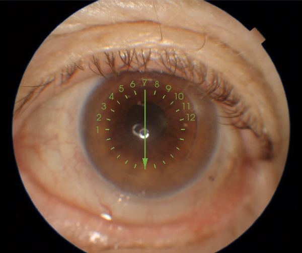
The final design seated, under the ptotic lid that displaced every lens before it.
Where he is now
He reads 20/25 with the lens and wears it about eleven hours a day with comfort maintained. At annual review the fit held: 360° clearance, excellent edges, no impingement. He has two spectacle corrections — a progressive worn over the scleral lens, and a separate pair for when the lens is not in.
The lens was replaced once, for warpage, surface scratches and increasing inferior decentration. The replacement showed the same mild inferior displacement that had characterised the fitting from the first lens onward.
That consistency is the lesson. When a lens decentres in the same direction across multiple unrelated geometries, the cause is unlikely to be the lens. In an eye like this one it is more realistic to aim for a fit that tolerates displacement — maintaining 360° clearance and acceptable edges despite it — than to chase perfect centration.
A handling incident, and what changed because of it
He attended an emergency department one evening after attempting removal with a plunger, several times, from an eye that contained no lens. The lens had been removed the previous day during a spectacle refraction and he had forgotten it was out. His wife had also attempted removal.
On examination the following day the graft was clear with inferior corneal staining, the anterior chamber was quiet, intraocular pressure was 12 mmHg and acuity was stable and correctable to 20/20. The impression recorded was inferior irritation. Topical antibiotic cover was prescribed and a colleague arranged to review him that week.
The counselling that followed is now routine for us with every plunger user: confirm the lens is present in the eye before any attempt at removal. It reads as obvious until it happens to a grafted eye.
Worth referring
Any post-keratoplasty patient with a clear graft and disappointing acuity — and any patient whose lid position is doing something a lens keeps having to answer for. If the graft itself is the limiting factor rather than the optics, we will say so and send them back.
See referring physicians for what comes back after a referral, or the other case studies.
One patient’s course, published with identifying details removed. Individual results depend on the graft, the ocular surface and the lids, and nothing here should be read as a typical or expected outcome. Dr. Shira Kresch is an optometrist, OD, MS, FAAO; keratoplasty is a surgical procedure performed by our surgical colleagues.