
Michigan's Contact Lens Specialists
Neurotrophic Keratitis Treatment in Michigan
Specialty Scleral Lenses for Neurotrophic Keratitis
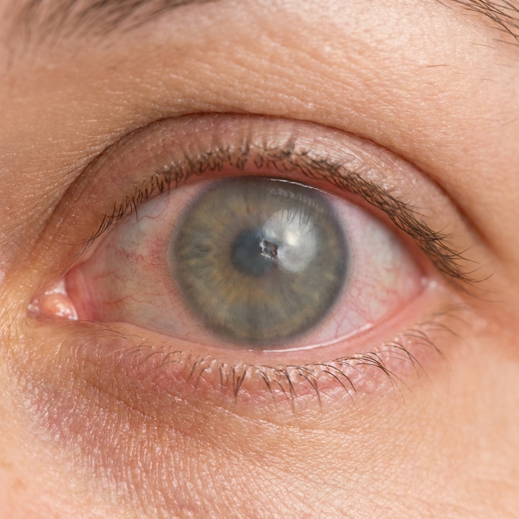
Neurotrophic keratitis — also called neurotrophic keratopathy, and sometimes neurotrophic keratoconjunctivitis or simply a neurotrophic cornea — is one of the most challenging corneal conditions to manage — and one where the right specialty scleral lens can make a profound difference. When the cornea loses its sensation, it also loses its ability to protect and heal itself. At Michigan Contact Lens, Dr. Shira Kresch fits protective scleral lenses that hydrate, shield, and stabilize a vulnerable cornea while your medical treatment addresses the underlying cause.
What Is Neurotrophic Keratitis?
Neurotrophic keratitis is a degenerative disease of the cornea caused by damage to the trigeminal nerve — the nerve responsible for corneal sensation. When the cornea can no longer feel, it loses the protective reflexes that keep it healthy: blinking, tear production, and the natural drive to heal minor injuries.
The result is a surface that gradually breaks down, developing persistent epithelial defects, ulcers, and — if left untreated — scarring and permanent vision loss. Because the cornea cannot signal pain, NK often progresses silently, which is why specialized monitoring and protection are so important.
What Causes Neurotrophic Keratitis and Am I at Risk?
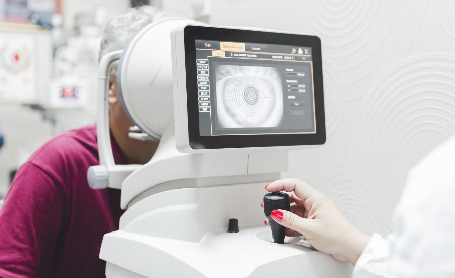
Neurotrophic keratitis develops when the corneal nerves are damaged or interrupted. Common causes include:
- Herpes simplex or herpes zoster (shingles) eye infections
- Diabetes and other conditions affecting nerve health
- Prior eye or brain surgery, including procedures near the trigeminal nerve
- Refractive surgery. LASIK and PRK cut corneal nerves by design and sensation normally recovers over months. Where recovery is incomplete, the result is sometimes called LASIK-induced neurotrophic epitheliopathy — worth investigating properly rather than managing as ordinary post-surgical dry eye
- Chemical or thermal burns to the eye
- Long-term contact lens overwear or chronic ocular surface disease
If you have any of these risk factors and notice changes in your vision or comfort, a specialized corneal evaluation is worthwhile — even if your eye doesn’t hurt.
Recognizing the Symptoms of Neurotrophic Keratitis
Because reduced sensation is a hallmark of the disease, symptoms can be deceptively mild. Watch for:
- Reduced corneal sensation — the eye feels numb and injuries go unnoticed
- Persistent redness or irritation — often with surprisingly little pain
- Blurred or declining vision — as the corneal surface becomes irregular
- Slow-healing surface defects — wounds that won’t close on their own
Corneal Sensitivity: the Test That Makes the Diagnosis
Everything about this disease follows from one measurement: corneal sensation.
Reduced sensation is called corneal hypoesthesia; complete loss is corneal anaesthesia. A healthy cornea is the most densely innervated tissue in the body, and those nerves do two jobs. They trigger the protective reflexes — blinking, reflex tearing, the instinct to close the eye — and they release factors that keep the surface epithelium healthy and healing. Lose the nerve supply and you lose both at once.
Testing it is quick and painless. A fine wisp of cotton drawn to the cornea, or a calibrated aesthesiometer, shows within seconds whether the reflex is intact. We test corneal sensitivity as a routine part of a specialty evaluation rather than only when neurotrophic disease is already suspected — because by the time it is suspected, damage has usually already happened.
If your eye looks inflamed or has a surface defect and it does not hurt the way it should, that discrepancy is the finding. In a cornea with normal sensation, that degree of damage would be extremely painful.
How Neurotrophic Keratopathy Is Staged
Clinicians stage the disease by what the corneal surface is doing. The Mackie classification is the one most commonly used, and it matters because the stage determines how urgent protection becomes:
- Stage 1 — epithelial changes. The surface is irregular, dry in patches, and stains with dye, but is still intact. Reduced sensation is present and often the only other clue.
- Stage 2 — persistent epithelial defect. An area of surface has broken down and will not close. The edges become smooth and rolled, which is characteristic — a defect that has stopped trying to heal.
- Stage 3 — corneal ulcer and stromal involvement. The damage extends into the deeper layers. This is the stage where a neurotrophic corneal ulcer can progress to melting or perforation, and where protecting the surface becomes urgent rather than optional.
Because the eye cannot signal pain, a patient can move between these stages without noticing. That is the argument for scheduled monitoring rather than symptom-driven visits.
Medical Treatment, and Where Lenses Fit
Neurotrophic keratitis treatment runs on two tracks at once: address the nerve and the surface medically, and protect the cornea mechanically while that works. We co-manage the first and provide the second.
On the medical side, the options your corneal specialist may use include:
- Preservative-free lubrication, frequently, and stopping anything toxic to the epithelium — which often means reviewing drops that are being used with good intentions but are making the surface worse.
- Autologous serum eye drops, made from your own blood serum, which carry growth factors that commercial tears do not.
- Cenegermin, a recombinant human nerve growth factor approved specifically for neurotrophic keratitis — the first treatment aimed at the nerve deficit itself rather than only its consequences.
- Amniotic membrane, applied to promote healing of a defect that will not close.
- Surgical options in advanced disease, including tarsorrhaphy to partially close the lids and corneal neurotisation to restore nerve supply.
None of those protects the cornea from a blink, from air, or from drying out between doses. That is what the lens is for, and it is why the two approaches belong together rather than competing.
How Scleral Lenses Treat Neurotrophic Keratitis
For a cornea that can’t protect or heal itself, a scleral lens does the work the eye no longer can. The lens vaults completely over the cornea and holds a reservoir of sterile fluid against the surface throughout the day.
- A continuous fluid reservoir keeps the cornea constantly hydrated
- The lens acts as a physical shield against blinking friction and airborne debris
- It creates a stable, protected environment that supports healing
- Vision becomes sharp and stable over an otherwise irregular surface
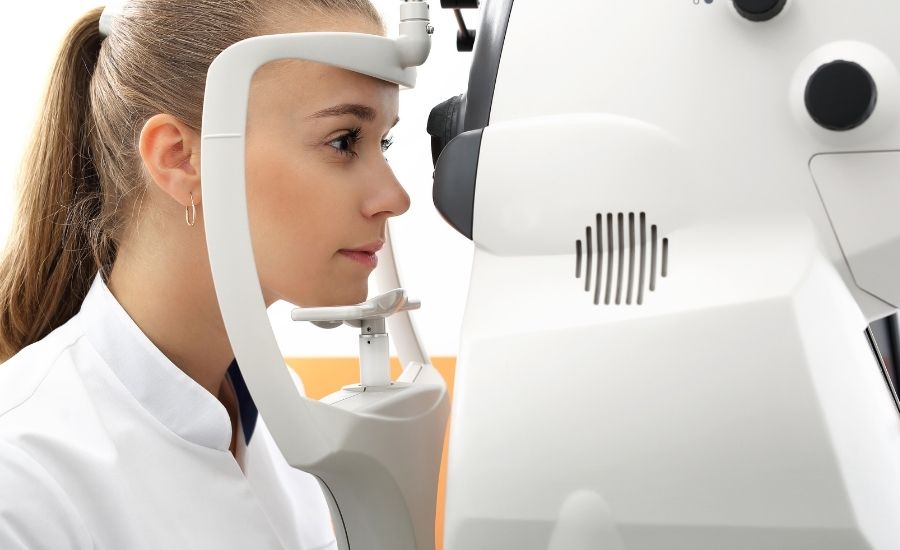
- Each lens is custom-mapped to your eye with Eaglet Eye profilometry
Scleral lenses are used alongside medical therapy — they protect and stabilize the eye while the underlying nerve condition is treated.
Bandage & Protective Lens Options
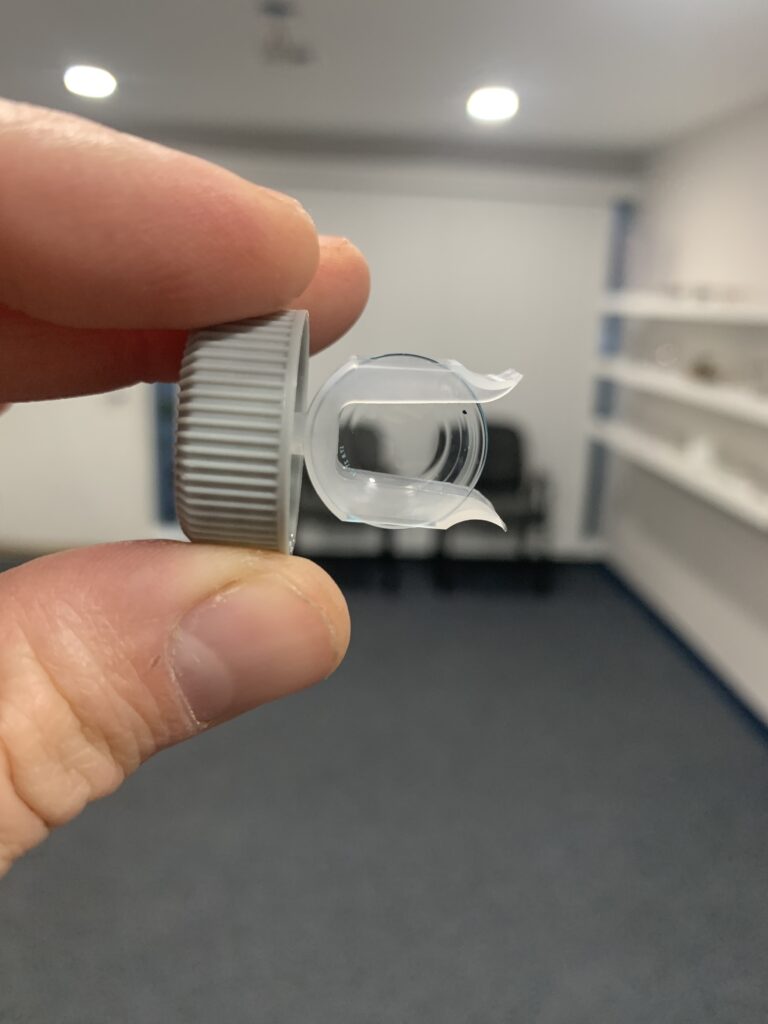
In earlier or milder cases, or between stages of treatment, other protective lenses may play a role. Bandage soft lenses can temporarily cover and protect a healing surface, while specialty designs bridge the gap until a full scleral lens is the right fit. Dr. Kresch selects the safest option for the current health of your cornea.
Working With Your Corneal Specialist
Neurotrophic keratitis is best managed as a team. Michigan Contact Lens co-manages NK with ophthalmologists and corneal surgeons across Metro Detroit, coordinating lens care around your medical treatment — whether that’s medication, amniotic membrane therapy, or surgical intervention. Early, coordinated care offers the best chance of protecting your vision.
Neurotrophic Keratitis FAQs
- Can neurotrophic keratitis be cured?
The underlying nerve damage is managed rather than fully cured, but the disease can be controlled and your vision protected. Scleral lenses play a major role by shielding and hydrating the cornea while medical therapy addresses the cause.
- Are scleral lenses safe for a damaged cornea?
Yes. Because scleral lenses vault over the cornea and never touch it directly, they’re one of the safest options for a fragile surface. The fluid reservoir actively protects and hydrates the eye all day.
- Do you work with my corneal specialist?
Absolutely. We co-manage neurotrophic keratitis with ophthalmologists and corneal surgeons, coordinating lens care around your medical treatment plan.
- Will my vision come back?
Many patients regain significant clarity once the surface is protected and stabilized. Outcomes depend on how much scarring has already occurred, which is why early specialized care matters.
- Is neurotrophic keratitis the same as neurotrophic keratopathy?
Yes — the two names describe the same disease. "Keratitis" emphasises the inflammatory component and "keratopathy" the degenerative one, and clinicians use both. You may also see neurotrophic keratoconjunctivitis, or simply a neurotrophic cornea. The condition is the same: corneal damage that follows loss of corneal nerve sensation.
- What is corneal hypoesthesia?
Corneal hypoesthesia means reduced corneal sensation, and complete loss is called anaesthesia. It is the defining feature of neurotrophic disease and the reason the cornea stops protecting itself. It is also easy to test: a fine wisp of cotton or a calibrated aesthesiometer touched to the cornea shows immediately whether the reflex is intact, and we test it routinely rather than only when we suspect a problem.
- How is neurotrophic keratitis treated?
Treatment runs on two tracks. Medical therapy addresses the nerve and the surface — preservative-free lubrication, stopping anything toxic to the epithelium, autologous serum drops, cenegermin (a nerve growth factor approved for this condition), amniotic membrane, and surgical options such as tarsorrhaphy or corneal neurotisation in advanced disease. Alongside that, a scleral lens protects and hydrates the cornea continuously while it heals. The two are complementary, not alternatives.
- Can LASIK cause neurotrophic keratitis?
Refractive surgery cuts corneal nerves by design, and sensation usually recovers over months. In a minority of patients recovery is incomplete, producing what is sometimes called LASIK-induced neurotrophic epitheliopathy — persistent dryness and a surface that heals poorly, with reduced sensation on testing. It is worth investigating properly rather than treating as ordinary post-surgical dry eye.
- Why does my eye look bad but not hurt?
That combination is the hallmark of neurotrophic disease, and it is the reason it is so often caught late. Pain is the cornea's alarm system, and in neurotrophic disease the alarm is disconnected. A red eye with a visible surface defect and little or no discomfort should raise the question immediately — in a healthy cornea, that degree of damage would be extremely painful.