
CASE STUDY · SJÖGREN’S SYNDROME
Past the last drop
A woman in her sixties, thirty years since diagnosis, who had worked through the entire medical ladder and was still symptomatic by lunchtime.
What had already been tried
Her own summary was that she had tried everything else, and the record supported it. Before she reached us she had been through autologous serum eye drops, punctal plugs, amniotic membrane, topical ciclosporin, a nasal secretagogue spray, lubricant ointment, lid hygiene and preservative-free artificial tears used throughout the day. She was still on several of these at presentation.
This is the position a Sjögren’s patient reaches when the disease is severe: not untreated, but maximally treated and still limited.
The argument for a scleral lens here is mechanical, not pharmacological. Every item on that list delivers an intermittent intervention to a surface whose deficit is continuous. Drops last minutes against a problem that runs all day. A scleral lens holds a reservoir of preservative-free saline against the cornea for the whole wearing period, so the surface is bathed rather than periodically wetted. That is a difference in mechanism, which is why it can work where the ladder has been exhausted.
The complication that shaped the fitting
Slit lamp examination found mild blepharitis with capped meibomian glands and an instantaneous tear break-up time in both eyes — expected in this disease. It also found conjunctivochalasis, and that changed the landing zone strategy.
Redundant conjunctiva can be drawn beneath the lens edge and prolapse into the post-lens reservoir. The usual instinct in haptic design is to minimise landing zone compression and avoid blanching. Here the opposite was done deliberately: the superior landing zone was tightened, and mid-peripheral blanching of the superior vessels was accepted as the intended consequence rather than treated as an error. The trade-off was recorded at the time. No significant conjunctival prolapse occurred at any point.
A caveat worth stating, because the literature does not fully support the simple version. Published work attributes conjunctival prolapse primarily to limbal clearance and lens settling rather than to landing zone compression, and reports prolapse in a substantial minority of scleral lens wearers. The two accounts reconcile — a tighter landing zone resists settling, and settling is the proximate driver — but a single case cannot establish which mechanism operated. What it supports is narrower: with conjunctivochalasis, the target is not simply “no blanching”, and the decision should be made consciously and written down.
Why the first lenses did not work
The first rotationally symmetric lenses produced a characteristic set of failures. The right lens rotated, with the orientation marking displaced, and decentred slightly inferiorly. The left decentred infero-temporally with touch along the full nasal vertical rim. Later, focal impingement was documented over a small inferior vessel on the right and a temporal vessel on the left.
She could wear them, but a pattern emerged: after about five hours she became aware of the lens edges, the right developed suction that made removal difficult, and both eyes ached and reddened after removal. Notably, during those five hours she needed markedly less supplementary lubrication — the reservoir was doing its work while the fitting relationship was not yet right.
Flattening the right lens by one step did not resolve the removal difficulty. That is the diagnostic detail. Scleral shape is asymmetric in most eyes, and a rotationally symmetric haptic on an asymmetric sclera cannot be corrected by adjusting a single global sagittal depth, because the misfit is not global.
What resolved it
The design was changed to a bi-elevation prolate configuration, with the horizontal and vertical sagittal depths specified independently — a 250 µm differential in each eye. A plasma surface treatment was specified to support wettability on an aqueous-deficient surface. Rotation, decentration and impingement resolved together.
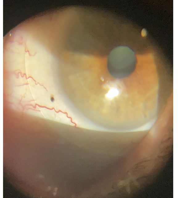
Her lens edge landing on the conjunctiva, vessels running underneath it undisturbed.
Presbyopia was addressed with monovision, which she described as mildly disorienting but preferable to distance-only, with no difficulty driving. She uses over-spectacles for closer work.
Where she is now
She wears the lenses around ten hours daily with comfort maintained throughout, reads 20/20 at distance in the dominant eye, and has settled into a maintenance routine: surfactant cleaning, a preservative-free saline rinse, a saline fill supplemented with autologous serum, a periodic deep clean, and a scheduled lens-free day every two weeks to rest the ocular surface. Fit assessments show 250–275 µm of central clearance, 360° clearance maintained, and no significant staining on removal.
Her own summary: the only relief is when the contacts are in.
The lens did not replace the dry eye regimen. Her serum concentration was escalated over time and a secretagogue added. What changed is that the regimen became tolerable and effective. Framing a scleral lens as a cure for Sjögren’s would misrepresent what happened here.
Worth referring
A Sjögren’s or severe ocular surface disease patient who has genuinely exhausted drops, plugs and serum. In our experience that point is reached earlier than it is acted on — these patients are often maintained on a ladder that has stopped helping because the next rung is assumed to be more of the same.
The lens treats the surface, not the disease, and sits alongside whatever rheumatology has in place. See referring physicians, or the other case studies.
One patient’s course, published with identifying details removed. Individual results depend on the cornea, the ocular surface and the underlying disease, and nothing here should be read as a typical or expected outcome. Dr. Shira Kresch is an optometrist, OD, MS, FAAO.