
Michigan's Contact Lens Specialists
Corneal Dystrophy Treatment in Michigan
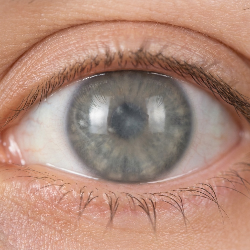
Clearer Vision With Corneal Dystrophy
Corneal dystrophies are inherited conditions that gradually cloud or distort the cornea, often affecting both eyes. As vision becomes hazy or irregular, everyday tasks get harder — but specialty lenses can help. At Michigan Contact Lens, Dr. Shira Kresch fits custom scleral lenses that smooth an irregular surface and restore clearer, more comfortable vision for patients with corneal dystrophy.
What Are Corneal Dystrophies?
Corneal dystrophies are a group of mostly inherited disorders in which abnormal material builds up in one or more layers of the cornea. They typically affect both eyes, progress slowly, and are not caused by injury or outside factors.
There are many types, grouped by the corneal layer they affect — including Fuchs’ dystrophy (innermost layer), map-dot-fingerprint dystrophy (surface layer), and lattice or granular dystrophies (middle layer). Each affects clarity and comfort differently.
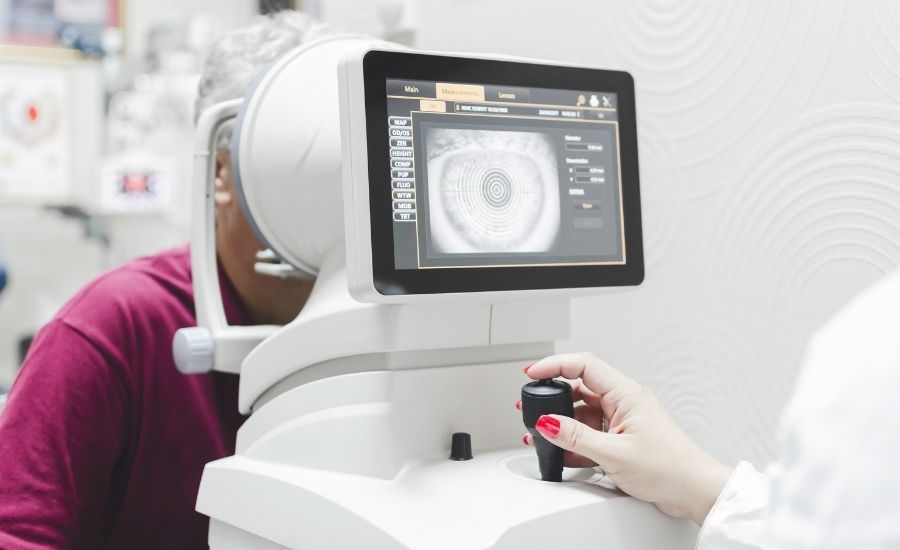
Types and Causes
Most corneal dystrophies are genetic and run in families. Common types include:
- Fuchs’ endothelial dystrophy — affects the cornea’s inner pump layer, causing swelling and haze
- Map-dot-fingerprint dystrophy — affects the surface, causing irregularity and erosions
- Lattice and granular dystrophies — deposits in the middle layer that cloud vision
Because they’re progressive, dystrophies benefit from ongoing monitoring by a corneal specialist.
Symptoms of Corneal Dystrophy
Symptoms vary by type but often include:
- Blurred or hazy vision , sometimes worse in the morning
- Glare and halos around lights
- Recurrent painful corneal erosions
- Light sensitivity
- Vision that fluctuates or isn’t fully corrected by glasses
How Scleral Lenses Help Corneal Dystrophy
When a dystrophy makes the corneal surface irregular, a scleral lens can restore clarity that glasses can’t reach.
- The lens creates a smooth optical surface over an irregular cornea
- A fluid reservoir hydrates the eye and eases recurrent erosions
- Vision becomes sharper, with less glare and haze
- The lens protects a fragile surface throughout the day
- Each lens is custom-mapped with Eaglet Eye profilometry
For surface and mid-layer dystrophies especially, scleral lenses offer meaningful comfort and visual improvement.
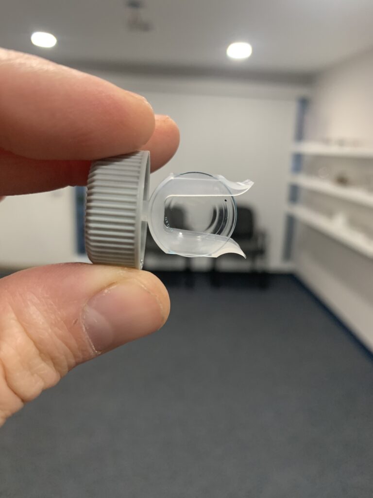
Fuchs’ dystrophy, in more detail
Fuchs’ is the most common corneal dystrophy and the one most people arrive having already been told they have, so it is worth setting out properly.
The innermost layer of the cornea — the endothelium — works as a pump, continuously drawing fluid out of the corneal tissue to keep it clear. In Fuchs’, those cells die off progressively and are not replaced, because human corneal endothelial cells do not meaningfully regenerate. As the pump weakens, fluid accumulates and the cornea swells and hazes.
The hallmark symptom is vision that is worse on waking and improves through the morning. Overnight, with the lids closed, fluid cannot evaporate from the corneal surface, so swelling peaks by morning. Once the eyes are open, evaporation gradually clears some of it and vision improves over an hour or two. If that pattern sounds familiar, mention it specifically — it is one of the more useful things a patient can report, and it points at the endothelium rather than the surface.
On examination the tell-tale sign is guttae, small bumps on the innermost membrane, often visible well before symptoms begin.
As it advances, swelling can reach the surface layer and form fluid blisters called bullae. When those rupture they are genuinely painful and expose the eye to infection. That stage is uncomfortable rather than dangerous in itself, but it needs managing rather than enduring.
Treatment usually runs in stages: hypertonic saline drops or ointment to draw fluid out osmotically, then, when vision or comfort no longer holds up, a partial-thickness endothelial transplant — DMEK or DSAEK — which replaces the failed pump layer rather than the whole cornea. Those are surgical decisions, made with a corneal surgeon.
Where a scleral lens fits in Fuchs’ — and where it does not
Worth being direct here, because this is the point where a specialty lens practice can easily oversell.
A scleral lens does not treat corneal oedema. It does nothing about the endothelial pump, and swelling is what drives the vision loss in Fuchs’. If oedema is the limiting factor, a lens will not fix it, and we will say so.
What a lens can do is address the other problem. Guttae and surface changes scatter light and produce glare and irregularity that glasses cannot correct, and a scleral lens gives those eyes a smooth refracting surface. Where painful epithelial bullae are present, a lens can also protect the surface in the way a bandage lens does.
Both of those come with a condition: the endothelium in a Fuchs’ cornea is already compromised, so the fit is designed conservatively and monitored more closely than a standard scleral fitting. Wearing time is built up gradually, corneal thickness is tracked, and we co-manage with your corneal specialist rather than working around them.
The honest summary is that in Fuchs’ a scleral lens is sometimes useful, always carefully considered, and never a substitute for the surgical conversation when the pump has failed.
Map-dot-fingerprint dystrophy and recurrent erosions
This one affects the outermost layer, where the epithelium is poorly anchored to the tissue beneath.
The characteristic history is sharp pain on first opening the eyes in the morning — during sleep the lid can adhere to the loose epithelium and pull a patch of it away on waking. Episodes come and go over years, often with long quiet spells between, and in between them vision may be near normal or slightly irregular.
Here a scleral lens has a clearer role than in Fuchs’. The fluid reservoir keeps the surface continuously hydrated and shielded through the wearing day, which can substantially reduce erosion episodes, and the lens smooths the optical irregularity at the same time.
Lattice and granular dystrophies
These deposit abnormal material in the middle layer of the cornea, gradually clouding it. Vision declines slowly and glare increases.
A lens helps with the irregularity and the glare, but as with any opacity, it cannot see through deposits — where they sit in the visual axis they cap what any correction can deliver. Something worth knowing about these specifically: they can recur in a graft years after a transplant, which is one reason a lens is often worth exhausting first.
When to seek care promptly
Most dystrophy symptoms change slowly, over years. These do not, and are worth acting on:
- Sudden sharp pain on waking, especially with watering and light sensitivity — a possible erosion
- A rapid drop in vision over days
- Redness with pain and discharge, which needs ruling out as an infection the same day
Slow change is the expected pattern. Fast change is not.
When Lenses Aren’t Enough
Some dystrophies — particularly advanced Fuchs’ — may eventually require surgical treatment such as a partial corneal transplant. Even then, scleral lenses can play a role before and after surgery. Dr. Kresch will help you understand where lenses fit in your long-term plan.
Working With Your Corneal Specialist
Corneal dystrophies are best followed over time. Michigan Contact Lens co-manages dystrophy patients with ophthalmologists and corneal surgeons across Metro Detroit, providing specialty lens care that complements your medical monitoring and any surgical treatment.
Corneal Dystrophy FAQs
- Can scleral lenses help corneal dystrophy?
For dystrophies that make the corneal surface irregular — map-dot-fingerprint, lattice, granular — yes. A scleral lens creates a smooth optical surface and keeps the eye hydrated, improving both clarity and comfort. For endothelial dystrophies such as Fuchs’, the picture is more nuanced: a lens addresses glare and irregularity but does nothing about the corneal swelling that usually drives the vision loss.
- What is the first sign of Fuchs' dystrophy?
Typically vision that is blurry on waking and clears over the first hour or two of the day. Overnight, with the lids closed, fluid cannot evaporate from the cornea, so swelling peaks by morning and then improves. Before symptoms begin, an examination may show guttae — small bumps on the innermost corneal membrane — which is often how it is picked up first.
- Can a scleral lens make Fuchs' dystrophy worse?
It is a genuine consideration rather than a theoretical one, which is why these fittings are approached conservatively. The endothelium in a Fuchs’ cornea is already compromised, so we design the fit to avoid excessive vault, build wearing time up gradually, track corneal thickness through the process, and co-manage with your corneal specialist. If oedema rather than surface irregularity is limiting your vision, we will tell you that a lens is not the answer.
- Are corneal dystrophies hereditary?
Most are. They are typically inherited and affect both eyes, which is why family history and regular monitoring by a corneal specialist matter. If a parent or sibling has been diagnosed, it is reasonable to have your own corneas examined even without symptoms.
- Will I need a corneal transplant?
Not necessarily. Many patients manage well with specialty lenses for years. Advanced Fuchs’ may eventually need a partial-thickness endothelial transplant — DMEK or DSAEK — which replaces the failed pump layer rather than the whole cornea. Scleral lenses can still have a role before and after that surgery.
- Do scleral lenses help with painful erosions?
Often, yes, and this is where they are most clearly useful. In map-dot-fingerprint dystrophy the epithelium is poorly anchored and can tear on waking. A scleral lens keeps the surface hydrated and shielded through the day, which can reduce how often erosions happen.