

Receiving a diagnosis of Keratoconus can be unsettling. This progressive eye condition, which causes the cornea to thin and bulge into a cone shape, can lead to significant vision distortion that glasses can’t always fix. The journey ahead can feel uncertain, and the most important first step is finding the right expert to guide your care.
Your search for a top keratoconus doctor in Michigan ends here. At Michigan Contact Lens, our clinic is a center of excellence for the diagnosis and management of keratoconus. We understand the unique challenges this condition presents and are dedicated to using the most advanced technology and treatments to protect and restore your vision.
This comprehensive guide will walk you through everything you need to know about keratoconus, from early symptoms and diagnosis to the full spectrum of modern treatment options. We’ll show you why choosing a true specialist is critical and how our team provides the expert care you need.
Keratoconus is a progressive eye condition that affects the cornea, the clear, dome-shaped surface of the eye. In a healthy eye, the cornea has a round shape, but in keratoconus, the cornea thins and begins to bulge outward into a cone shape. This structural change prevents light from focusing correctly on the retina, leading to significant visual challenges.
This condition typically begins in the teenage years or early 20s and can worsen over time if not managed properly by a qualified keratoconus doctor in Michigan. According to the National Keratoconus Foundation, it affects approximately 1 in every 2,000 individuals. Left untreated, keratoconus can severely impact vision and quality of life, making tasks like driving at night or reading difficult and frustrating.
The exact cause of keratoconus is not fully understood, but it is believed to be linked to a combination of genetic, environmental, and biochemical factors. A keratoconus specialist in Michigan will consider all these factors during your evaluation.
Genetics play a significant role. About 1 in 10 people with keratoconus have a parent who also has the condition. It is also more common in individuals with certain systemic conditions like Down syndrome, Ehlers-Danlos syndrome, and other connective tissue disorders.
There is a very strong association between chronic, vigorous eye rubbing and the progression of keratoconus. The physical stress and friction from rubbing can weaken the already compromised corneal structure. This is often linked to conditions that cause itchy eyes, such as allergies, atopic dermatitis, or eczema.
The cornea is primarily made of interwoven collagen fibers that give it strength and shape. In keratoconus, these fibers are weaker. It is believed that an imbalance in enzymes within the cornea leads to oxidative damage, which further weakens the collagen and allows the cornea to bulge.
The symptoms of keratoconus can be subtle at first but typically worsen as the cornea becomes more irregular. It’s crucial to see a doctor if you experience any of the common signs of keratoconus.
This is the primary symptom. It’s not the same as normal nearsightedness. Patients often describe it as looking through a warped or wavy window. Straight lines may appear bent or crooked.
The irregular cone shape of the cornea scatters light as it enters the eye instead of focusing it cleanly. This causes disabling glare from sources like headlights, streetlights, and even computer screens, making night driving particularly difficult.
One of the earliest red flags for a developing keratoconus is a rapid and frequent change in an eyeglass prescription, especially a significant increase in astigmatism. If your vision seems to be getting noticeably worse every year, it warrants a specialist evaluation.
This is a hallmark symptom of keratoconus. It’s the perception of a faint, transparent second image trailing the primary one, like a comet’s tail. If you cover one eye and still see double or multiple images, it’s a clear sign of a corneal irregularity.
The brain works incredibly hard to try and make sense of the poor-quality, distorted images it’s receiving from the affected eye. This extra effort can lead to persistent eye strain and headaches.
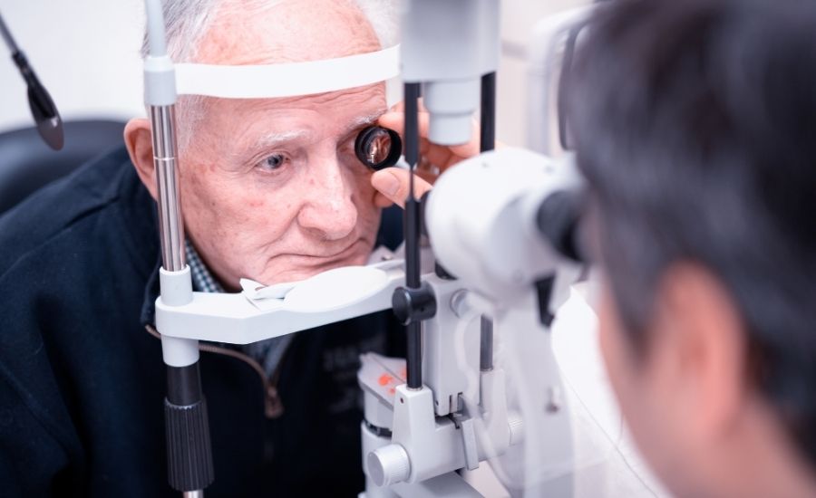
A definitive diagnosis is the first and most important step. A comprehensive eye exam by our keratoconus doctor in Michigan will include several painless, advanced tests:
Early detection is crucial for effective management, allowing for interventions that can halt the progression of the disease.

The right keratoconus treatment in Michigan depends on the stage and progression of the condition. Our goal is always twofold: first, stop the condition from getting worse, and second, provide the best possible vision.
Corneal Cross-Linking (CXL) is a revolutionary, minimally invasive procedure and the only treatment proven to stop keratoconus from getting worse. It is a vital early intervention to preserve the cornea’s integrity.
The procedure involves applying riboflavin (Vitamin B2) eye drops to the cornea, which are then activated by a controlled UV light. This process creates strong new bonds between the collagen fibers in the cornea, making it stronger and more stable. As noted by the American Academy of Ophthalmology, CXL is highly effective at preventing the need for future, more invasive surgery. We work with the top surgeons for corneal cross linking in Michigan to co-manage your care.
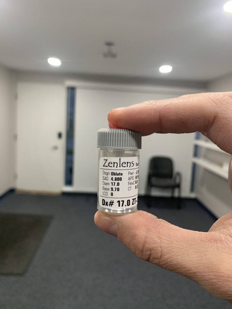
For restoring clear vision, the premier non-surgical keratoconus treatment is a custom-designed scleral lens. These large-diameter lenses vault completely over the irregular cornea, creating a perfectly smooth optical surface. The space between the lens and the eye is filled with saline, neutralizing all distortions.
The fitting process for these lenses is complex and requires advanced technology and expertise. However, the result is often life-changing, providing a level of comfort and clarity that no other non-surgical option can match. Our clinic is a leading provider of scleral lenses for keratoconus in Michigan.
In some mild or moderate cases, smaller Rigid Gas Permeable (RGP) lenses or hybrid lenses (with a hard center and soft skirt) can also be effective at improving vision. Our specialists will determine the best option for your specific corneal shape.
For the most advanced cases where the cornea is significantly scarred or contact lenses are no longer effective, surgical intervention may be required. This can include a partial-thickness transplant (DALK) or a full-thickness corneal transplant (PKP). Our keratoconus doctor in Michigan will co-manage your care with the best corneal surgeons in the state.
At Michigan Contact Lens, our keratoconus doctor in Michigan will often recommend scleral lenses as the premier vision-correcting option for individuals with moderate to advanced keratoconus. These lenses rest on the stable, less sensitive sclera (the white of the eye), completely vaulting over the irregular cornea.
The space between the lens and the cornea is filled with sterile saline, which creates a perfectly smooth new optical surface and enhances comfort by constantly bathing the eye in moisture. The custom fit of scleral lenses for keratoconus in Michigan provides the clarity of gas-permeable lenses with the comfort of soft lenses, ensuring stability and minimal movement during blinks.
This makes them a superior option for managing keratoconus compared to traditional lenses, and fitting them correctly is a hallmark of the best keratoconus doctor in Michigan.
In most cases, yes — keratoconus is a medical diagnosis, not a routine vision issue, so medically necessary treatments like corneal cross-linking and specialty contact lenses (including sclerals) are often covered by medical insurance plans rather than vision plans. Coverage specifics vary widely by carrier and plan. Our team helps you navigate your benefits, provides any necessary documentation, and supplies a superbill for out-of-network reimbursement when applicable.
No — LASIK is strictly contraindicated for patients with keratoconus. The procedure removes corneal tissue to reshape the cornea, which would dangerously weaken an already thinned and compromised keratoconic eye and could trigger rapid progression. This is one of the most important reasons to see a keratoconus specialist before any refractive surgery. A thorough corneal topography screening can rule out subclinical keratoconus that might not show up on standard exams.
Follow-up frequency depends on disease activity. Patients with actively progressing keratoconus are typically seen every 3 to 6 months to monitor corneal changes and consider cross-linking. Patients with stable disease in well-fitting scleral lenses are usually seen annually. After cross-linking, patients return at 1 month, 3 months, 6 months, and 12 months to confirm the treatment held and the cornea remains stable.
The earliest signs are often subtle and easy to miss. Common early symptoms include frequently changing eyeglass prescriptions (especially worsening astigmatism), blurred or distorted vision that glasses no longer fully correct, increased light sensitivity and glare, ghosted or double vision in one eye, and eye rubbing that makes vision temporarily worse. Keratoconus typically begins in the teens or twenties and progresses for 10 to 20 years before stabilizing.
No — keratoconus does not cause complete blindness. However, untreated advanced keratoconus can severely impair functional vision, making daily tasks like driving, reading, or recognizing faces very difficult. The good news is that modern treatment is highly effective: corneal cross-linking halts progression in most cases, and properly fit scleral lenses restore excellent vision even in advanced disease. Early diagnosis and treatment produce the best long-term outcomes.
There is a clear genetic component. About 1 in 10 keratoconus patients have an affected family member, and genome studies have identified several genes associated with corneal thinning. If a parent, sibling, or child has keratoconus, you should have your corneas screened with topography — even if you currently have good vision. Other risk factors include chronic eye rubbing, atopic conditions like eczema and asthma, and connective tissue disorders.
Yes, and pediatric keratoconus tends to progress faster and more aggressively than adult-onset cases — which is why early diagnosis matters so much. Children with allergies who rub their eyes frequently, kids with Down syndrome, and those with connective tissue disorders are at higher risk. If your child has rapidly changing prescriptions, especially worsening astigmatism, ask their eye doctor to perform corneal topography. Cross-linking is FDA-approved for patients age 14 and up.
Keratoconus is a specific progressive corneal thinning that causes the cornea to bulge into a cone shape. It’s often confused with other conditions: pellucid marginal degeneration (PMD) thins the lower cornea differently; post-LASIK ectasia is a similar weakening caused by refractive surgery; keratoglobus involves more diffuse corneal thinning. Corneal topography distinguishes between them, and the distinction matters because treatment approaches differ significantly.
Don’t let the uncertainty of a keratoconus diagnosis control your life. With modern treatments and expert care, clear, comfortable vision is achievable. The first step is a consultation with a true specialist.
Contact Michigan Contact Lens today to schedule your comprehensive evaluation. Let our team show you the path to better vision.
Make an appointment on WhatsApp