
If glasses or regular contacts no longer give you clear, comfortable vision, scleral lenses may be the right next step. These custom lenses rest on the white of the eye and hold a cushion of fluid over the cornea, improving vision, comfort and stability through the day.
This guide covers what scleral lenses are, the different types and sizes, who they help, what fitting involves, how to care for them, the problems people run into, and when a symptom means you should stop wearing them and call your doctor.
Michigan Contact Lens fits scleral lenses for patients across Metro Detroit — Southfield, Troy, Birmingham, Royal Oak, Farmington Hills and Detroit — for dry eye, keratoconus, irregular corneas and other hard-to-fit eyes.
Scleral lenses — also called sclera lenses or sclera contacts — are large-diameter rigid gas-permeable lenses. Unlike a standard contact lens, which sits directly on the cornea, a scleral lens arches over the cornea and lands on tissue over the sclera, the white of the eye.
That single design difference is what makes them work. Three things follow from it:
They are designed to vault the cornea. The fit is checked after settling to confirm adequate clearance. For an eye that is scarred, irregular, thinned or hypersensitive, the aim is that nothing bears on the damaged surface — which is why sclerals are often tolerated by patients who cannot wear any other lens. Clearance is not fixed: the lens settles into the tissue over the first hours of wear, so vault is assessed on the eye rather than assumed from the design.
They hold a fluid reservoir. The space between the back of the lens and the front of the eye is filled with preservative-free saline before insertion. The cornea sits in liquid all day. For selected patients with severe ocular surface disease, the fluid reservoir can provide substantial protection and symptom relief.
They replace the corneal surface optically. Light entering the eye meets the smooth front surface of the lens rather than an irregular cornea. The rigid front surface and fluid reservoir reduce the optical effect of corneal irregularity. This is why vision in keratoconus is often much better in a scleral than in glasses — though some residual higher-order aberration can remain, which is what wavefront-guided designs are aimed at.
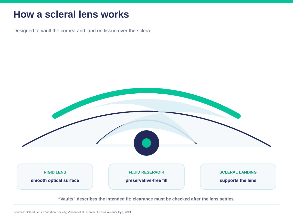
See it for yourself: our interactive scleral lens simulator shows how the lens vaults the cornea and where the fluid reservoir sits.
Scleral lenses are made from rigid gas-permeable polymers — not glass, and not the soft hydrogel used in disposable lenses.
“Rigid” here describes optical behaviour, not comfort. The material holds its shape, which is precisely why it can replace an irregular corneal surface: a soft lens would drape over the irregularity and reproduce it. Because the lens rests on the sclera, which carries far fewer nerve endings than the cornea, most patients find a scleral more comfortable than a small rigid lens, and many find it more comfortable than soft lenses.
The critical property of the material is oxygen permeability, usually written as Dk. The cornea has no blood supply and draws oxygen directly from the air. A scleral lens plus its fluid reservoir sits between the cornea and that air supply, so the material has to let oxygen through. Modern high-Dk polymers are what made large-diameter lenses viable — earlier materials could not transmit enough oxygen at these diameters and thicknesses.
This is a point where sclerals are often underestimated. The materials used for them are among the most oxygen-permeable in contact lens manufacturing — comfortably more permeable than conventional soft hydrogels, and at the top end matching or exceeding the best silicone hydrogels. A lens being rigid and thick does not make it a low-oxygen lens.
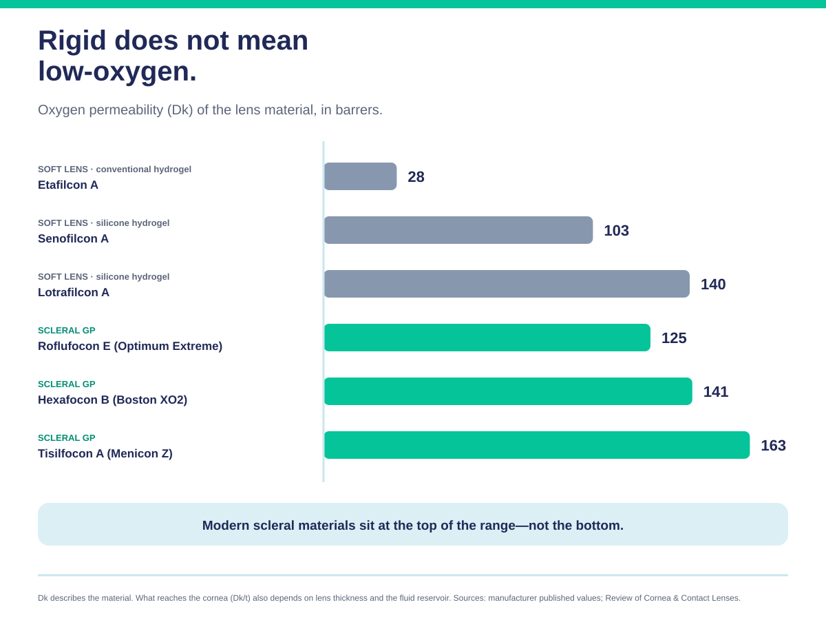
Two design factors interact with this. Oxygen has to cross the lens and the fluid reservoir behind it, so in principle a thicker lens or a deeper reservoir slows delivery. Early theoretical models set fairly strict limits on both. Clinical studies since have been more reassuring: measured corneal swelling has not tracked reservoir depth the way those models predicted across the range normally fitted, and making lenses thinner has not reliably reduced it either. The practical conclusion is that a high-Dk material matters more than chasing the shallowest possible vault — which is useful, because vault depth also has to satisfy clearance, settling and landing, and those cannot be traded away.
Different laboratories use different proprietary materials and designs, which is why a lens that suits one patient is not simply transferable to another. A guide to scleral lens brands →
“Scleral lens” describes a family of lenses, not one product.
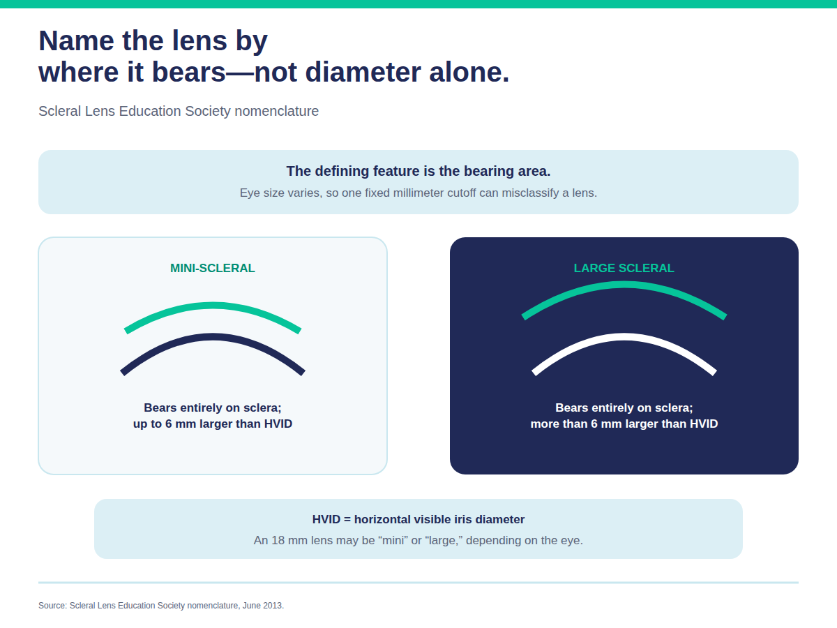
The names are often assumed to be diameters, and that is the wrong way round. The Scleral Lens Education Society classifies these lenses by where they bear — which part of the eye carries the lens — and sizes them relative to your eye rather than to a universal millimetre cutoff. The reference point is the horizontal visible iris diameter (HVID), roughly the width of the coloured part of your eye, which varies from person to person.
That is why the same physical lens can be called different things on different eyes. An 18 mm lens may be a mini-scleral on one patient and a large scleral on another. Diameter ranges quoted by laboratories are rough manufacturing conventions, not definitions.
Mini-scleral lenses
Bear entirely on tissue over the sclera, up to about 6 mm larger than your HVID. They clear the cornea entirely but stay small enough to be relatively easy to handle. For many patients, a mini-scleral is the practical middle ground. More on mini-sclerals →
Large scleral lenses
Bear entirely on tissue over the sclera, more than about 6 mm larger than your HVID. A larger lens changes where and how the lens lands, and can open up fitting strategies that a smaller lens cannot — which is often useful in advanced keratoconus, significant corneal ectasia and severe ocular surface disease.
A common misconception is worth correcting here: a bigger lens is not automatically a deeper one. How much fluid sits over your cornea is set by the lens’s sagittal depth, its design and the shape of your eye — not by diameter on its own.
Impression-molded and profilometry-designed lenses
Rather than fitting from a standard set, the lens is designed from a three-dimensional map or physical impression of your eye’s surface. This matters when the sclera itself is asymmetric — which it commonly is — because a lens designed to the real shape lands more evenly. Our profilometry-designed approach →
Wavefront-guided scleral lenses
These are designed to reduce selected higher-order aberrations that a conventional lens cannot address. Relevant for patients still troubled by glare, halos or ghosting even after a good conventional fit. Wavefront-guided lenses →
Multifocal scleral lenses
Add near correction for presbyopic patients who also need a scleral. Multifocal sclerals →
Prosthetic (tinted) scleral lenses
A scleral lens made with a tinted iris and pupil, used to restore appearance after injury or disfigurement and sometimes to reduce glare where the iris is damaged. This is a distinct thing from an ocular prosthesis (an artificial eye, fitted by an ocularist for an eye that has been removed or has no vision) and from PROSE (a separate prosthetic device design fitted through its own network of trained providers). The three are frequently conflated online and should not be blended here. Prosthetic sclerals →
Which type suits you is a fitting decision, not a shopping decision — it depends on your corneal shape, scleral shape, condition and handling ability. How a custom scleral lens is designed and fitted →
Scleral lenses are used where the cornea is irregular, the ocular surface is compromised, or standard lenses cannot be worn comfortably.
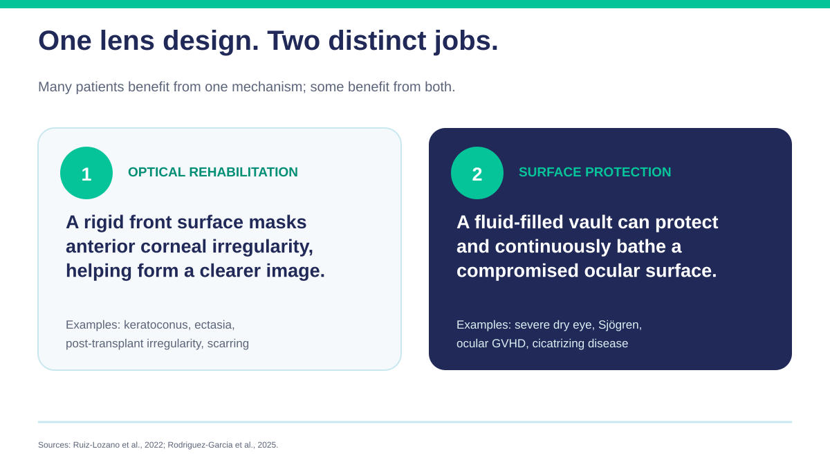
There are two broad reasons a scleral helps, and they are worth separating, because they explain why one lens type serves such different patients.
Reason one — it replaces the optical surface. If the cornea is misshapen, a scleral gives light a smooth surface to enter through instead.
Reason two — it protects and hydrates. If the ocular surface is diseased, the lens shelters it and keeps it in fluid all day.
Some patients need one. Many need both.
Keratoconus — one of the most common reasons patients are referred to us for a specialty lens. The cornea thins and bulges into a cone, and the resulting irregular astigmatism often cannot be fully corrected in glasses, which address regular curvature. A scleral vaults the cone. Many patients describe the first fitting as the clearest their vision has been in years.
Pellucid marginal degeneration — a thinning disorder affecting the lower cornea. The irregularity sits peripherally, which makes it particularly awkward for smaller lenses that must land near it. The larger diameter of a scleral is an advantage here.
Corneal ectasia and irregular corneas after LASIK or PRK — where a cornea has weakened and distorted after refractive surgery. These eyes are often both irregular and dry, so both mechanisms apply.
After a corneal transplant — a graft heals with a junction and often leaves significant astigmatism that glasses cannot correct well. A scleral can vault the graft and the junction rather than bearing on them. Timing matters: fitting over a graft is not routine, and when it is appropriate depends on how far healing has progressed, whether sutures remain, and the health of the graft’s endothelium. We fit these eyes in coordination with your cornea surgeon, only once they have cleared you for lens wear, and with closer monitoring than a standard fit.
Corneal scarring from injury or infection — scarring creates a fixed irregularity that glasses often cannot fully correct.
Irregular astigmatism — astigmatism that is not a simple regular curve, and therefore often not fully correctable in spectacles.
Severe dry eye that has not responded to drops, punctal plugs or other treatment. The reservoir means the cornea is bathed continuously rather than between drops, which for patients instilling drops through the day can make a substantial difference to comfort.
Sjögren’s syndrome — an autoimmune condition affecting tear-producing glands. Because tear production itself is impaired, a scleral lens can provide a continuously hydrated environment for the ocular surface.
Graft-versus-host disease — ocular GVHD after stem cell transplant can produce severe surface disease. Sclerals are a recognised part of management.
Stevens-Johnson syndrome — severe surface and eyelid damage where protection from the lids’ own movement matters as much as hydration.
Exposure-related surface disease — where the eye does not fully close or the surface is chronically exposed.
Patients who have tried soft or rigid lenses and could not wear them — because of dryness, instability, discomfort, or lenses that would not stay centred. This is a common referral: not a diagnosis in itself, but a history of everything else having failed. More on hard-to-fit contact lenses →
Not everyone with these conditions needs a scleral lens, and not everyone with one of these diagnoses is a candidate. Who is a good candidate? →
A successful scleral fit starts with the right expertise. We combine advanced corneal and scleral measurement, specialty fitting experience, and structured follow-up for patients with complex corneal conditions and contact lens intolerance.
We use advanced corneal mapping to design lenses around your exact eye shape — a more precise fit, better day-to-day comfort, and more reliable vision, particularly for patients who have not done well elsewhere.
Dr. Shira Kresch, OD, MS, FAAO, has advanced training in corneal disease and specialty lens design. Patients travel from Southfield, Troy, Birmingham, Royal Oak, Farmington Hills, Detroit and across Metro Detroit for scleral lens fitting.
Scleral lenses are not fitted from a standard set of sizes. Each fit is built for your eye.
1. Consultation and ocular health evaluation. We establish or confirm the diagnosis, review what you have already tried and why it failed, and examine the health of the ocular surface. This last part matters: an eye with active inflammation or untreated lid disease will not do well in any lens until that is addressed, so sometimes the first step is treatment rather than fitting.
2. Corneal and scleral mapping. Imaging captures the shape of the cornea — and, importantly, the sclera. This is where fitting has changed most in recent years. The sclera is not a sphere. It is typically asymmetric, and the pattern of that asymmetry differs between individuals and between the two eyes of the same person. A lens designed to a real measured surface lands more evenly than one selected from a standard set. Our profilometry-designed approach →
3. Diagnostic lens fitting. You wear trial lenses so the fit can be assessed on your eye rather than on paper. We are looking at four things:
4. Design refinement. Parameters are adjusted — overall diameter, vault depth, limbal zone, landing-zone geometry, and power — and the lens is ordered from a laboratory that manufactures to those specifications. Because these are individually lathed rather than picked from stock, there is a manufacturing interval between visits.
5. Dispensing and training. We teach insertion, removal, filling and care, and you do not leave until you can handle the lenses yourself. This session is longer than people expect and is the part that determines whether the fit succeeds in real life.
6. Follow-up. The lens rests on living tissue for the whole day, so follow-up is part of the fit, not an optional extra. We check what the lens is doing after hours of wear — settling, reservoir clarity, how the landing zone looks on removal — because a lens that looks ideal at dispensing can behave differently after eight hours.
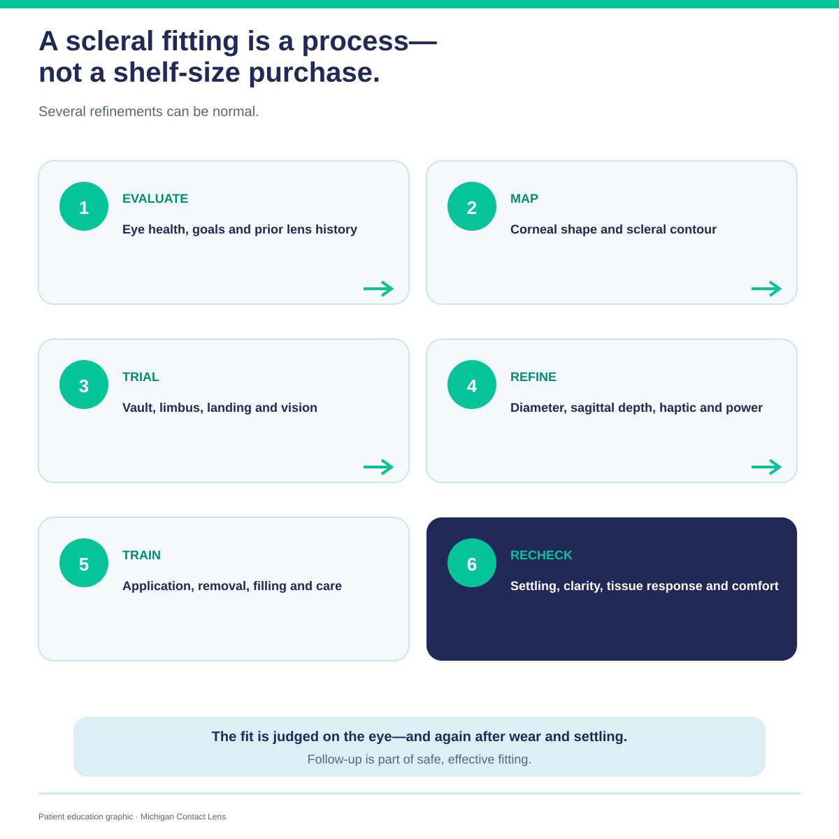
Because these lenses are highly customised, more than one visit is normal, and a fit that takes several refinements is not a fit that has gone wrong. What to expect at your fitting →
Most patients are comfortable quickly, but there is a learning curve.
Handling. Insertion and removal take practice. Handling usually becomes easier with practice, but the learning curve varies — some patients are comfortable within days, others take considerably longer, and needing more training time is not a sign the lens is wrong for you. Insertion and removal guide →
Vision settling. Vision can shift slightly during the first hour of wear as the lens settles into the tissue. This is expected. Why vision shifts →
Building wear time. We usually build wear time up gradually rather than jumping to full days. Wearing schedule →
Comfort. Most patients report the lenses feel better than what they wore before. Adapting in the first two weeks →
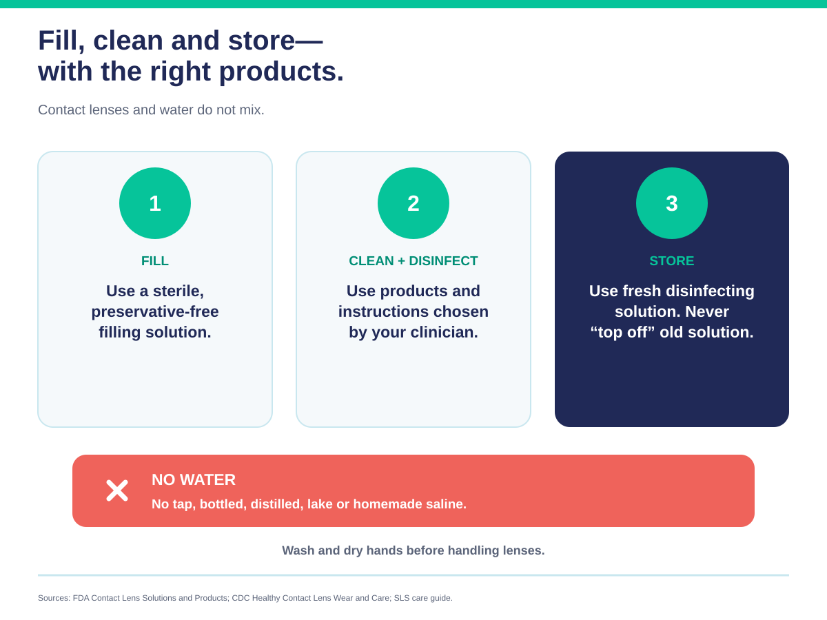
Scleral lenses are filled with fluid and worn against a compromised eye. Care is not optional.
Filling. Fill with sterile preservative-free saline. Never use water of any kind — not tap, not bottled, not distilled, and not homemade saline. Water carries organisms, including Acanthamoeba, that can cause a severe and sight-threatening corneal infection. Saline options →
Cleaning. Clean after every removal with a solution recommended for rigid lenses. Care, cleaning and solutions →
Storage. Store in fresh disinfecting solution. Never top off old solution.
Drops. Not every eye drop is compatible with a scleral in place. Compatible drops →
Handling aids. A plunger makes removal easier and is standard equipment. Using a plunger →
Dr. Kresch and Dr. Melissa Barnett wrote the guide we wish every patient had on day one — insertion and removal step by step, troubleshooting, supplies and daily care, in far more detail than a web page allows.
Download the free guide →Midday fogging. Vision goes hazy after several hours. Two separate things cause it: debris collecting in the fluid reservoir behind the lens, or the front surface of the lens clouding up — which is usually driven by dry eye and meibomian gland dysfunction. Sometimes the fit is contributing as well. Why fogging happens →
The lens will not come out. Removal difficulty has several possible causes — technique, the lens adhering after a long day, or a landing zone that is fitting too tightly — and it is not something to diagnose yourself. Do not force it. A drop or two of preservative-free artificial tears in the eye before you try again will often loosen the seal — that is the first step the Survival Guide gives too. Then use the removal technique and plunger you were trained on; if that still does not work, contact us rather than persisting. Recurrent difficulty means the fit needs reviewing. If a lens is stuck →
Air bubbles under the lens. Most often a filling or application issue — not enough fluid in the bowl, or the lens tilting as it goes on. Recurrent bubbles despite good technique can indicate a fit problem instead, so mention it if it keeps happening. Air bubbles →
A red ring after removal. An impression where the lens landed. A faint mark that fades is common; a persistent or angry ring means the landing zone needs adjustment. Red ring →
Cloudy lenses. Several things can cause this: deposits on the lens surface from skin oils or makeup, poor surface wettability, particulate matter getting into the reservoir, an interaction with a solution you are using, or the fit itself. Frequent cloudiness is worth investigating rather than living with.
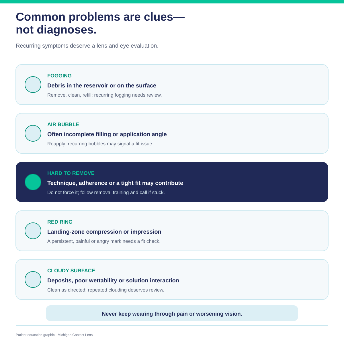
Most of these are solvable. None should be simply tolerated — if something recurs daily, the lens needs looking at.
Scleral lenses are not right for everyone, and they ask more of the patient than a standard lens.
The fitting takes time. Several visits, sometimes over weeks. This is not a same-day product.
They cost more than standard contacts. Custom design, laboratory manufacture and extended follow-up all contribute. What determines the cost →
Handling is a real skill. Patients with significant tremor, very limited dexterity or difficulty raising their arms may struggle. This is worth discussing honestly before starting.
They require commitment to care. The fluid reservoir means poor hygiene carries real risk, not just discomfort.
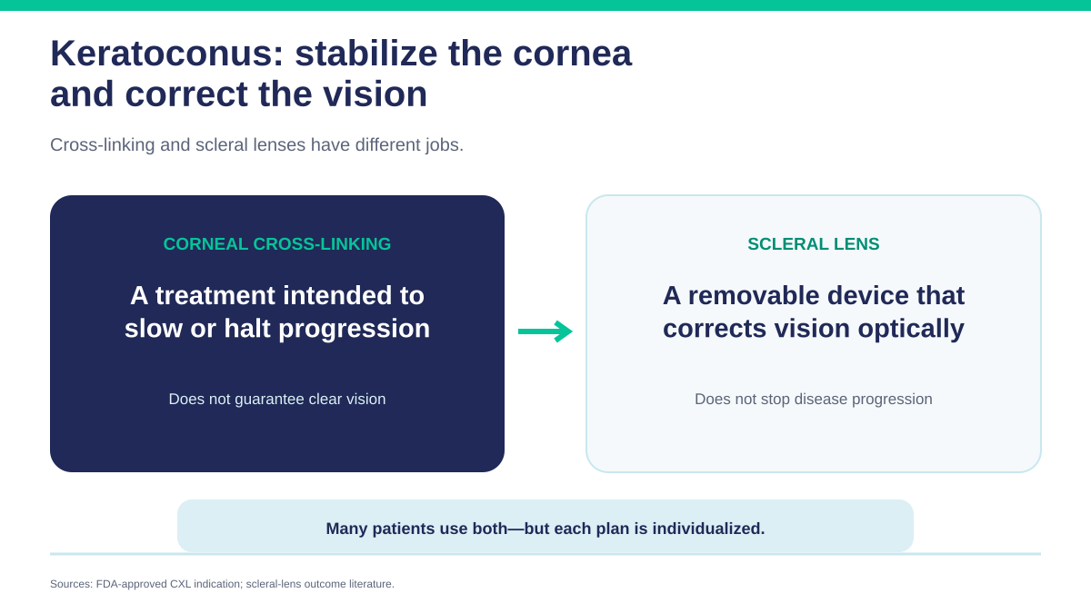
They do not treat the underlying condition. A scleral lens manages keratoconus optically — it does not halt progression. Cross-linking is the treatment that addresses progression, and the two are often used together. Scleral lenses after cross-linking →
Some eyes are not suitable. Certain scleral shapes, surface conditions or previous surgeries make a successful fit unlikely. We would rather tell you that at the consultation than after several attempts.
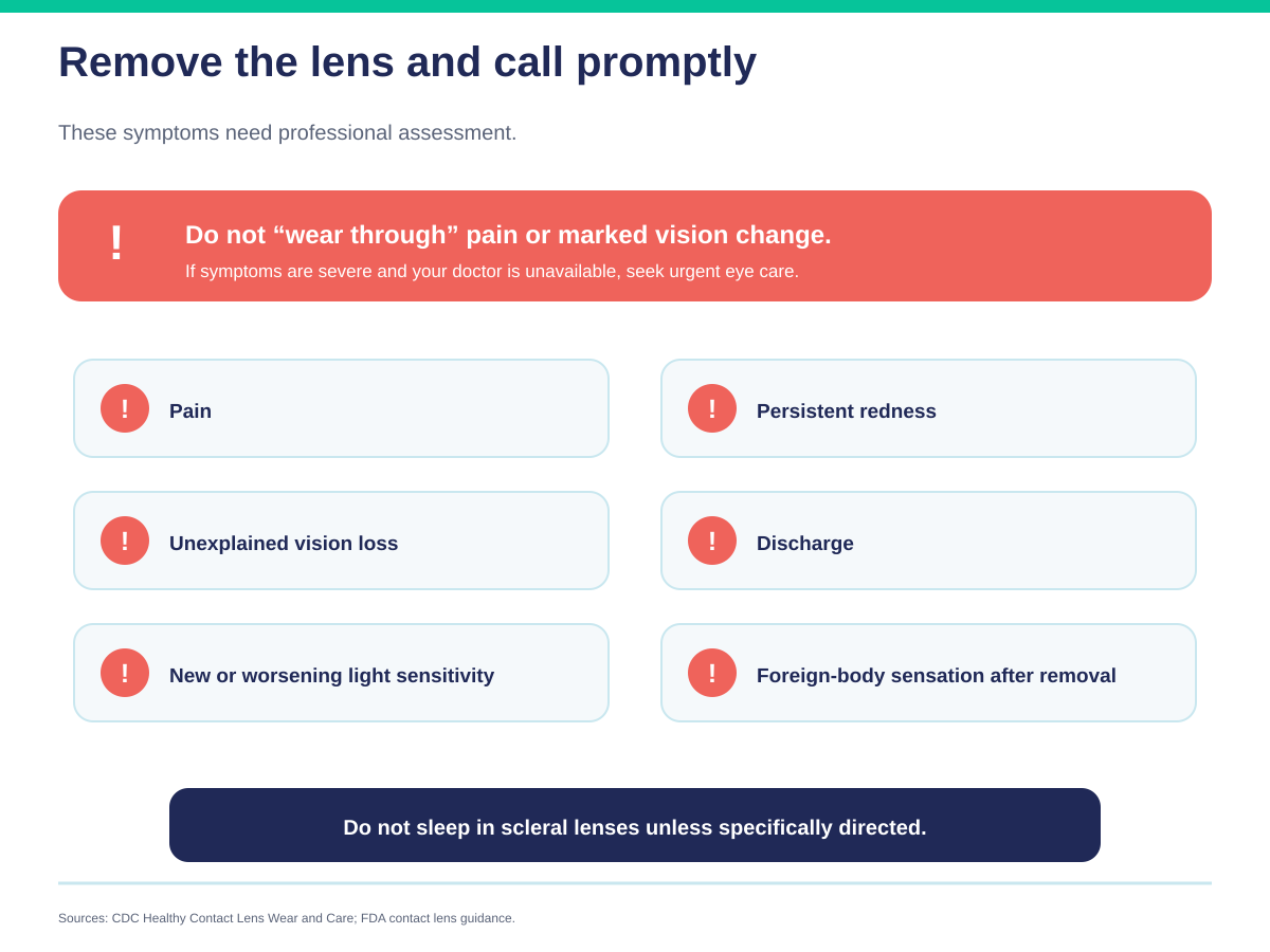
Remove the lens and contact your eye doctor promptly if you experience any of the following:
These can indicate corneal swelling, an abrasion or an infection. Prompt assessment is important because infection and corneal injury can threaten vision.
If you cannot reach us and symptoms are severe — significant pain, marked vision loss — seek urgent eye care rather than waiting.
Do not wear the lenses while sleeping unless we have specifically instructed you to.
Clearer, more stable vision; better comfort; and a secure fit for eyes that are difficult to fit. They reduce the optical effect of corneal irregularity, tend to stay centred more consistently than smaller lenses, and hold a fluid cushion that helps with dryness and sensitivity. For many patients, this is the first lens that is both clear and genuinely wearable.
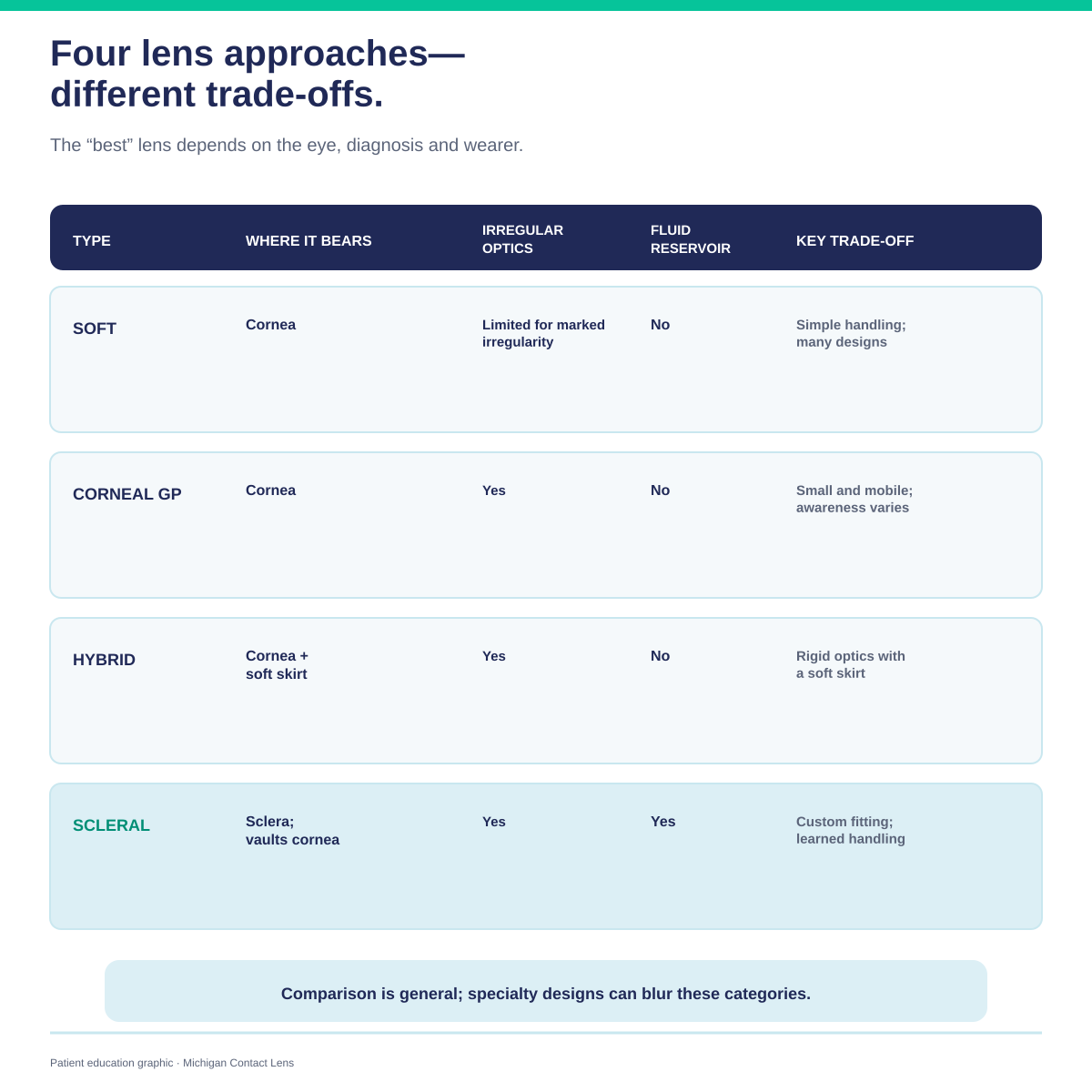
These are general tendencies, not fixed properties. Lens design has moved a long way — there are specialty soft designs made specifically for irregular corneas, and materials and geometries vary widely within each category. Which lens suits you is decided on your eye, not from a table.
| Where it bears | Irregular-cornea optics | Fluid reservoir | Typical trade-offs | |
|---|---|---|---|---|
| Soft lens | On the cornea | Standard designs tend to drape over irregularity and reproduce it; specialty designs for irregular corneas exist | None | Vision can be unstable on an irregular cornea |
| Corneal RGP | On the cornea | Good — a rigid front surface | None | Lens awareness; can dislodge; often less well tolerated on a sensitive or dry eye |
| Hybrid | Rigid centre on the cornea, soft skirt beyond it | Good | None | Aims to combine RGP optics with soft comfort; handling and replacement differ from either |
| Scleral | Entirely on tissue over the sclera, vaulting the cornea | Good | Yes — continuous | Costs more; longer fitting; handling is a learned skill |
Versus soft lenses. A standard soft lens takes on the shape of what it covers, so on an irregular cornea much of the irregularity comes through. That is the usual reason patients with keratoconus move off standard soft lenses — though specialty soft designs for irregular corneas are an option for some eyes and are worth asking about.
Versus corneal RGP lenses. A small rigid lens also gives a smooth optical surface, and for some eyes it is the right answer — it is simpler and less expensive. But it rests on the cornea itself, which is why it is less comfortable on a sensitive or diseased eye and more likely to dislodge. Sclerals vs RGP →
Versus hybrid lenses. A rigid optical centre with a soft skirt — an attempt to combine RGP optics with soft-lens comfort. A reasonable middle option for some patients. Hybrids for keratoconus →
Versus corneal transplant. This is the comparison that matters most to patients with advanced keratoconus. For some people with keratoconus, successful scleral-lens wear may help defer or avoid keratoplasty — retrospective studies have found lower transplant rates in populations where scleral lenses became widely available, which is an association rather than proof that a lens prevents surgery. What can be said plainly is that a lens is reversible and avoids the risks that come with surgery — though lens wear carries its own risks, which is what the two sections above are about — and that it corrects vision without treating the disease. This is a decision to make jointly with your cornea specialist, not one to settle from a web page. Sclerals vs transplant →
Many lenses are replaced around every one to two years, but timing depends on your condition, the fit, your prescription, the surface quality of the lens and your clinician’s guidance. One study at a tertiary-care centre found a mean replacement interval of about 24 months, with wide variation between patients — which is the honest summary: there is a typical range, and individual lenses fall well either side of it.
In practice, replacement is driven by surface scratching, deposits that no longer clean off, a change in your prescription, or a change in the eye itself. How long scleral lenses last →
Cost varies with your prescription, the complexity of the fit and the follow-up required. The value is in the evaluation, the custom design and a lens that genuinely works for your eye. A factor-by-factor breakdown →
We are an out-of-network practice. Our Insurance & Payment Options page explains superbill reimbursement, HSA/FSA and CareCredit.